Tag Archives: equine biomechanics

Hilary Clayton:  Biomechanical Interactions Amongst the Rider, the Tack and the Horse

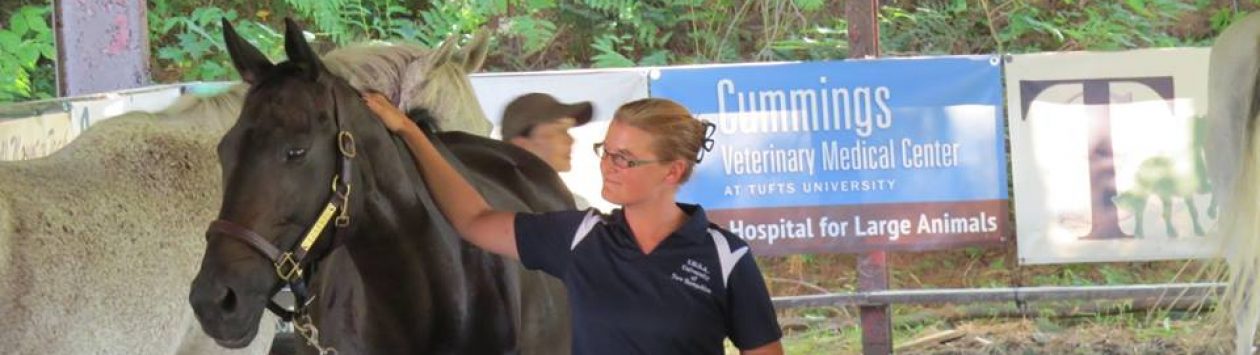
Dr. Clayton shares her thoughts at the University of New Hampshire Equine Program on October 27, 2015

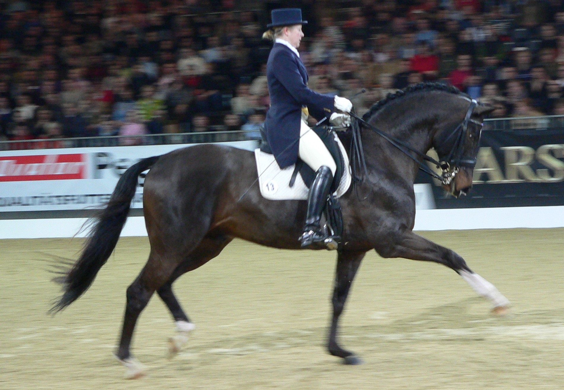
There is no doubt that how we ride our horses, the tack we use on them and the manner in which the horses carry themselves has a cumulative effect on their well-being over time.  Whether that effect is positive or negative is one of the questions considered in the study of equine biomechanics, which combines the disciplines of physics and physiology to study how forces and work affect the body of the horse.  Dr. Hilary Clayton is well known for the work she has done in this field, and she visited the University of New Hampshire in October of 2015 to share her thoughts on some of the more common interactions which occur amongst the horse, the rider and the tack.

HilaryClaytonPortrait
Dr. Hilary Clayton (Photo taken from her promotional poster.)

Equine Topline Mechanics 101

Clayton started her presentation with an overview of the structure of a horse’s vertebral column and how it works.  Just like in humans, the horse’s spine is made up of bone, ligaments, muscles and discs; equine discs are relatively thin compared to a human’s, and therefore horses don’t suffer from slipped discs and disc pain like humans can.  Even though the horse’s spine is horizontal, it loads similarly to a human’s in that it compresses together when force is applied.

Arabian_horse_skeleton
In this skeleton of an Arabian, it is easy to see the difference in the bones of the cervical and thoracic spine.  Note also the proximity of the spinous processes.

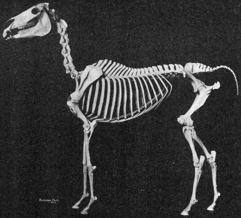
Each intervertebral joint has a small degree of mobility; when taken in totality, this allows for considerable movement along the entire length of the horse’s spine.  The degree and type of movement which the topline displays varies with each gait. In the walk, there is some bending and rotation in the topline, but little flexion or extension.  At the trot, there is more flexion and extension and the back is stabilized.  At the canter and the gallop, there is a great deal of flexion and extension, particularly in the lumbosacral joint, and the back is stabilized.

The Horse in Motion by Eadweard Muybridge

Instructors and trainers will often tell their clients that their horses need to move with a swinging back.  Clayton explained that this statement is not wholly accurate.  When in locomotion, the horse’s back must actually remain stable in order to support the horse’s weight and to transmit propulsive forces from the limbs. Clayton says that when the horse moves at liberty, they are not actively moving their back.  The back movement we see is due to gravity, inertia and the propulsive forces of the hind leg. In fact, excessive mobility of the bones in the spine during motion is never the goal—it is the muscles which need to be supple into order to control the movement of the spine.

Charlotte_DUJARDIN_JEM2014
Charlotte Dujardin and the incomparable Valegro.  Photo by Florence Skowron, via Wikimedai Commons.

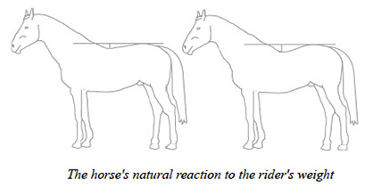
Clayton compared the structure of the articulated vertebrae of the horse’s spine to a beam which has a support at each end.  In the horse’s case, the “beam” tends to sag a bit in the middle, due to the weight of the internal organs and other viscera.  When we add the weight of a saddle and rider to this region, we increase the hollowing of the spine and the “dip” in the middle of the beam.  Clayton explains that with a rider, the range of flexion and extension is the same but the entire cycle of the motion is more extended.

Through the above description, it should be immediately apparent that the weight of the rider is inherently causing stress on the horse’s topline.  More troubling, though, is that when the horse’s back is hollowed as result of this weight, the dorsal spinous processes are approximated, which can lead to the development of the degenerative condition known as kissing spines.  Clayton’s research has shown that far more horses are affected with kissing spines than just those which show overt symptoms; however, even at a subclinical level, the syndrome can cause the horse discomfort and reduce the quality of their performance.  The good news is that when the back is rounded, the opposite effect occurs—the spinous processes are spread out. Therefore, regardless of your discipline, you horse should learn to work with a round topline.

kissingspines
In this image borrowed from a fellow wordpress user, the kissing spines can be clearly seen.

Round Backs and Development of the Horse’s Core

Clayton compared the mechanism which causes the horse’s back to be round to a “bow and string”.  The “string” is comprised of the muscles on the underside of the bones of the back, in this case the abdominal and sublumbar muscles.  The “bow” is therefore made up of the muscles located above the vertebrae.  Rounding their back requires the coordinated action of the horse’s core muscles.

Athletes of all species can achieve more optimal performance with a strong core.  These muscles are important both for balanced movement and coordinated stabilization.  Clayton divided the muscles of the horse’s core into three groups:  the back muscles, the sublumbar muscles and the abdominal muscles, and the groups work in concert to achieve the maximum mobility of the horse’s spine.

horsereactiontoweight
Image from http://www.equinechronicle.com

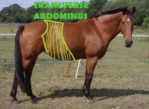
The back muscles make the topline hollow, round or bend right and left.  The sublumbar muscles flex the lumbosacral joint and the pelvis, which helps to bring the hind legs forward and underneath the body.  Finally, the abdominal muscles wrap all around the horse’s belly, running many different directions.  This group of muscles includes the transverse abdominal, the obliques and the rectus abdominus.  Collectively, they literally help hold the horse’s ‘guts’ in place, as well as stabilize the spine and assist with lateral bending.

Clayton explained the function of the back muscles in more specific detail. First, she discussed the longissimus and iliocostalis muscles, which are the long mobilizing muscles of the back.  They are made up of long fibers and cross many joints. These muscles are able to move the entire back of the horse.

The multifadi muscles serve the function of stabilizing the horse’s back.  These muscles are located right against the spinous processes and are comprised of short fibers which cross only a few joints; therefore, they work on only a limited area of the horse’s spine.  However, the condition of these muscles can have a profound effect on the shape of the back in a specific area.  More will be said on this later.

horse_anatomy_the_muscles_by_wiggle_chicken
Image from jenpenjen.deviantart.com

Clayton pointed out that most muscles work in pairs or layers; therefore, the deep stabilizing muscles are as important as the long mobilizing muscles, as they help to prevent vibration in the horse’s bones.  They also have a low activation threshold, which means that they will contract (along with the transverse abdominal muscle) simply in anticipation of locomotor activity.  They then serve to stabilize the horse’s spine as the limbs move.

Limited research has been done on the many effects of the horse’s stabilizing muscles on the spine.  However, research done on humans has shown that chronic back pain is often associated with atrophy of the deep stabilizing muscles, as joints then become too mobile.  Impaired spinal stabilization is an important risk factor and a predictor of recurrent back pain in humans.  Based on her research, Clayton extrapolates that a similar connection exists in horses.

core.stability.muscles
Notice how the same muscles provide stability to the human spine.  Image found on http://www.appliedpostureriding.com.au

Human research has also shown that even when back pain is resolved, the deep stabilizing muscles do not resume normal activity on their own.  Physiotherapy exercises are necessary to re-train the pre-activation of the stabilizing muscles.  In human patients who underwent this therapy, the one year recurrence rate of pain reduced from 80% to 30%.

When it comes to back pain, Clayton says that horses go through a similar cycle to humans.  When the back hurts, the deep stabilizing muscles become inactive, resulting in atrophy.  This causes the long mobilizing muscles to compensate, but since they are not equipped to stabilize the spine, these muscles spasm and cause further pain.  Therapeutic exercises are needed to reactivate the deep stabilizing muscles and to break the cycle of compensation and pain.

Developing Your Horse’s Core with Dynamic Mobilization Exercises

Clayton has developed a series of core strengthening exercises and sequences through her research, and she goes into illustrated depth about these in her book, Activate Your Horse’s Core (Sport Horse Publications, 2008). She gave a brief synopsis during her presentation.

Dynamic Mobilization Exercises are those in which the horse follows a controlled movement pattern which strengthens the muscles that move and stabilize the back.  They are comprised of rounding exercises and bending exercises.  In the rounding exercises, most of the flexion comes from the poll (high position) or the base of the neck (low position).  In the bending exercises, most of the movement comes at the base of the neck.

pilatesmobile1
Image from http://www.horseyard.com.au, from an article related to Dr. Clayton’s work.  

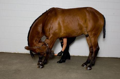
Clayton described a protocol which provided positive results in several “couch potato” school horses.  Using small bits of carrot to motivate the horse, they did three rounding exercises (chin to chest, chin between the carpi (knees) and chin between the fetlocks) and three lateral bending exercises (chin to girth, chin to hip, chin to hind fetlock).  For the lateral bending exercises, the human stood next to the horse, making them bend their neck around the human.  Horses did five repetitions/day and on left and right sides, if appropriate.  The exercises were repeated five days/week for three months.  Even with these stretches as the only form of exercise, the horses showed a positive development in their deep stabilizing muscles.

One of the benefits of these exercises is that the horse will only stretch as far as they are comfortable.  Ideally, the handler should encourage the horse to hold the stretch for as long as possible, but even stretching for a short period will help improve the strength of the multifidus muscle.   The best benefits are seen when these exercises are performed before the horse works each day; regular inclusion of them in a training program will help equine athletes throughout their career.  In addition, these exercises can be used in youngsters to help develop the deep stabilizing muscles before they begin under saddle training and are also especially beneficial for horses recovering from colic surgery, with appropriate approval from the attending veterinarian.

Transverse-Abdominus
Image from http://www.classicphysiotherapy.co.uk

The Role of Equipment and Rider in Equine Back Pain

When it comes to saddle selection and fit, it is clear that both the horse and the rider must be comfortable.  While the rider can simply vocalize their discomfort, the horse must express it in other ways, and it is important for riders and trainers to remain sensitive to this communication.  Unfortunately, the manner in which horses display the existence of back pain is as variable as the causes.  However, both the saddle and the rider can contribute to discomfort in the horse’s topline, and certain issues are almost sure predictors of pain in the horse.

horse-and-rider

Clayton has made extensive use of an electronic pressure mat which sits on the horse’s back in her research on saddles.  This specialized mat has 256 sensors (128 on each side of the spine) which measure force distribution on the horse’s back.  Her research has shown that the total force placed on the horse’s back varies with the size and weight of the rider and saddle as well as the gait of travel.

For example, in the trot, the suspension phase has minimal pressure, while the stance phase of each stride has the maximum force.  This is when the horse’s body is starting to rise up, but the weight of the rider is still down.  The mean force placed on the horse is at least equal to the rider’s weight in the walk; in the trot, it is two times the rider’s weight and in the canter it is three times.

equetrian-market-01
This image, taken from http://www.sensorprod.com, shows the pressure profile of a rider’s buttocks and thighs on the saddle.  Clayton’s tools offer her similar insight into the intensity, duration and location of pressure when a horse is in motion. 

Clayton says that she is frequently asked to quantify how much weight a given horse can fairly be asked to carry, but she says that this is a complex question to answer.  Variables such as the height, weight, conformation, fitness and soundness of the horse all play a role.  For example, a horse with a short and broad loin coupling can likely carry more weight than a horse of similar size with a long or narrow loin.  As far as the rider goes, variables such as weight, fitness, symmetry, balance, postural control and health issues all influence the impact they have on a given horse. Also important is the activity the horse is being asked to do—what type of work and on what kind of footing or terrain.

Finally, the saddle itself can have a positive or negative impact on a horse’s comfort level.  Each saddle is unique in terms of its load-bearing area, fit and suitability for a given horse, rider and job.   Soft tissues compress when pressure is applied.  The larger the area the force is spread over, the less overall pressure there will be.  This is one of the reasons why more modern saddles have long, broad panels.

Saddle-Panel-Diagram1-1024x501
Image from http://www.totalsaddlefit.com

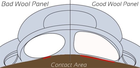
Pressure is calculated via force divided by area.  Therefore, the pressure increases if the force is larger or if the contact area is smaller.  Areas of deep pressure can be very harmful, causing ulcers or necrosis of the tissue due to increased capillary pressure.  On a less extreme level, pressure can cause discomfort through abrasions.  Clayton says that it is the magnitude and duration of pressure which are most important.  Muscles in particular are easily damaged by pressure.

If you see dry spots under your saddle after work, these are areas of increased pressure which prevent the sweat glands from working and are cause for concern.

dryspot
In this photo, it is clear where there has been an area of increased pressure and therefore inhibition of the sweating mechanism. 

Interesting Saddle Trivia

In her research, Clayton has had cause to investigate a number of different areas in which saddles might impact the horse, and she shared some of her findings with the audience.

One of the first questions she looked at was whether a saddle is truly necessary.  Most riders prefer to use a saddle for the stability and security it provides them on the horse’s back, and as it turns out, horses seem to prefer that their riders use saddles, too.  Without a saddle, the pressure of the rider is distributed over a smaller area, and the focal points of that pressure are over the rider’s seat bones.  (Interestingly, Clayton found similar results when she looked at one brand of treeless saddle, as well).  Clayton found that in general, a saddle which fits the shape of the horse’s back and the shape of the rider’s pelvis will provide stability to the rider’s position, and as a result, the pressure is more evenly distributed.  She also mentioned that within a breed, 80-90% of animals will have a similar back shape.

Correct saddle fit is of course of paramount importance.  Correctly fitted saddles are more stable, which increases horse and rider harmony.   The rigid parts of the tree, including the gullet plate, the points and the bars, can cause increased areas of pressure on the horse’s back.   It is important to consider the width of the gullet plate and length of the tree’s points in relation to the position of the scapula and related muscles.  When the horse extends their forelimb, the scapula rotates back and down on its back side, and rotates a little bit up in front.  This causes the back edge of the scapula to actually slide underneath the saddle in this moment of the stride.  A well fitting saddle should allow for free movement of the scapula when the forelimb is protracted.

Dia-2-Saddle-Fit
From http://www.equilibriumproducts.com

Clayton says that the width of the tree is equally important.  The correct width allows the load to be evenly distributed over a large area.  Ideally, the contact area is long and wide, with no focal points of high pressure.  A tree which is too wide may cause the gullet to put direct pressure on the withers and/or cause high pressure along the panels close to the spine.  In addition, the saddle often tips forward and down.  A tree which is too narrow is one of the most common causes of bridging; there is more pressure at the front and the back of the panels, and the saddle tips backwards.  Clayton says that bridging is the most common saddle fit problem, and it must be evaluated with the rider on board and while the horse is in motion.

bridging
Image from http://www.saddlemakers.org.  

More nuanced aspects of saddle fit include assessing the width of the gullet and slope and shape of the panels.  A wider gullet is usually better, because it allows mobility of the spine without causing it to hit the edge of the panels.  The slope of the panels must also suit the shape of the horse’s back. Panels come in a variety of widths and curvatures, and it is important that the type chosen suits the individual animal.

Finally, Clayton emphasized that saddle pads cannot compensate for the deficiencies of a poorly fitting saddle.  However, they may increase the horse’s comfort if the saddle is essentially the correct size and shape.  Clayton says that pads made with natural fibers, such as sheepskin, seem to have a better degree of resiliency and spring.

Girths and Slipping Saddles

Girth design has evolved considerably in recent years, and Clayton touched briefly on the subject at the end of her talk.  She said that the highest pressure beneath the girth occurs just behind the elbow in the moment when the forelimb contacts the ground. In her research, contoured girths seem to do the best job in terms of reducing both force and pressure.

Finally, Clayton discussed saddles which seem to constantly slip to one side.  She says that it is important to determine if the cause of the slip is the horse, the rider or the fit of the saddle.  Subtle hind limb lameness can be blamed for the cause of many slipping saddles, particularly when the slip occurs consistently to one side and with a variety of different riders on board.  In 60% of these cases, the saddle slips towards the side of the lame/more significantly lame hind limb.  The slip will go away when the lameness is eliminated through the use of nerve or joint blocks.  Clayton commented that rider crookedness is more likely to be an effect than the cause of saddle slipping.  Clearly if the cause of the saddle slip is lameness, this issue must be addressed before the problem will go away.

Final Thoughts

Clayton’s presentation covered a broad range of topics, but one theme was quite clear—riders have an obligation to their horses to ride them in as correct of a manner as possible, in the best fitting tack possible.  In this way, riders and trainers can actively contribute to the preservation of the horse’s long term soundness and promote their well-being.

Cadre_noir_-_saut_au_piquet.jpg
With good training and attention to correct tack and conditioning, horses are truly capable of amazing feats.  “Cadre-noir-saut au piquet” by Alain Laurioux


Hilary Clayton: It’s All About the Forelimb

Dr. Hilary Clayton: “It’s All About the Forelimb”

Presented at the USDF Convention 2014: Cambridge, MA

On December 5, 2014, I had the opportunity to attend my first ever USDF Convention, held in Cambridge, MA; my primary motivation for taking on the Friday AM commuter traffic to Boston was to hear a lecture being presented by Dr. Hilary Clayton.  Clayton (BVMS, PhD, Dipl ACVSMR, MRCVS) is truly a pioneer in the field of equine biomechanics and I have heard over and over that her lectures are not to be missed; it seemed silly to allow an opportunity to finally attend one to slip away.

Dr. Hilary Clayton   Photo taken from her promotional poster.
Dr. Hilary Clayton Photo taken from her promotional poster.

Clayton has written several books and is a frequent contributor to the USDF Connection; I enjoy reading her articles but I have always felt that some of her concepts go over my head.  Hearing her articulate and clarify her research was incredibly enlightening.  Here, I will attempt to summarize her remarks presented at the convention this year.  The section headings here mimic those of her talk; why reinvent them when her own words do such a good job?

The Limbs In General Terms

Clayton began her talk by explaining that the limbs of any species are made up of a series of rigid bones which articulate at moveable joints; these joints are stabilized and moved by muscles.  The length of the bones, combined with the angles of the joints, affect each limb’s ability to support body weight and/or provide propulsion to its owner.

If you think about a heavy species, such as an elephant, and you look at the skeletal structure of the limbs, you will see that they are strong, straight and vertical.  This design is excellent for bearing weight, but not so good for athletic endeavors.  Due to this structure, Clayton says that elephants are actually not capable of a moment of suspension and can’t jump, which is why a small moat will contain them at a zoo.  This is an example of a “limb as a supporting pillar”, according to Clayton.

Elephant limbs are post like and good for bearing weight, but not so good at creating propulsive force.

Species with small body weight, such as the cat, tend to have limbs with bones that are highly angled and joints which are compressed.  This allows a great deal of athleticism but sacrifices the ability to bear weight.  By opening up the angles of these compressed joints, these species are able to produce large amounts of propulsive force.  In addition, the spines of these species are usually more flexible, allowing two moments of suspension per stride in the gallop—once in flexion, and once in extension.  Clayton calls this anatomy “limbs as a propulsive lever”.

Cats are athletic and agile due to the angled nature of their limbs; however, their bodies could not support great weight.  By Les Chatfield from Brighton, England (Cat Skeleton  Uploaded by snowmanradio) [CC BY-2.0 (http://creativecommons.org/licenses/by/2.0)], via Wikimedia Commons
Cats are athletic and agile due to the angled nature of their limbs; however, their bodies could not support great weight. By Les Chatfield from Brighton, England (Cat Skeleton Uploaded by snowmanradio) [CC BY-2.0 (http://creativecommons.org/licenses/by/2.0)%5D, via Wikimedia Commons
The anatomy of the horse actually combines these two extremes:  the forelimb is more elephant- like and straight, while the hind limb is more cat-like and angled.

 In this skeleton of an Arabian, it is clear that the horse's front limbs are pillar-like while the  hind limbs are angled for propulsion.
In this skeleton of an Arabian, it is clear that the horse’s front limbs are pillar-like while the hind limbs are angled for propulsion.

In dressage and jumping training, the focus is correctly very much on the action and use of the horse’s hind limb, which ultimately controls the horse’s ability to leave the ground.  However, Clayton points out that “a fabulous hind leg is no good without an equally fabulous front limb.”  This is because the less angled joints and more upright posture of the horse’s front limbs allow them to act as struts for the horse; the forelimbs ultimately control the position of the forehand and most importantly, control the horse’s speeding and turning ability.

Ground Reaction Force

One of the coolest aspects of Clayton’s presentation was reviewing computerized footage of actual horses moving over the force plates at her former research facility at Michigan State University (Clayton became “emeritus” in April of 2014).   These videos showed how and where the force of movement translated itself through the horse’s body, and also how those vectors moved throughout the course of a stride.

Clayton explained the concept of Ground Reaction Force (GRF) as being the force which actually makes the horse move.  When the horse’s hoof is on the ground, it is automatically pushing against the ground; the GRF is the reaction of the ground pushing back against the hoof.  Because the front limbs bear more weight, the GRF is always higher on them.

The relative sizes and directions of the GRF’s of the forelimbs and hind limbs affect the horse’s balance.  Basically, it is the job of the hind limb to create propulsion, while the forelimb stops the horse’s balance from going wholly onto the forehand.  By changing the angle of the GRF, the horse controls his speed and direction.

To help us to understand the relationship between the GRF and the roles of the front and hind limb, Clayton used a video of a horse jumping a fence.  At take-off, the hindlimb forces cause the jumping horse to rotate forward, towards their center of balance.  The forward rotation is necessary for the horse to be able to take off from the hind limb and land on the fore limb.  At landing, the GRF of the forelimb causes a reversal in the direction of rotation, allowing the horse to shift back towards their center and land the hind limbs.

Finding the Balance

So basically, the horse’s body in movement is a set of opposing forces—one set from the hind limb which propels the horse forward, and one set from the fore limbs which prevent that force from pushing the horse down.  The conundrum is that the harder the hind limbs push and the longer they stay on the ground (so, increased engagement), the greater the tendency is for this force to rotate the horse onto the forehand.  When the hind limbs trail behind the horse, the force pushes the horse onto the forehand.  The role of the forelimbs becomes to maintain an uphill balance and counteract the tendency to fall forward.

Clayton reminded us that horses as a species have adapted to be “cursorial” (aka runners).  Cursorial species have limbs with certain qualities; in particular, the weight of the limb is concentrated in the upper section, with heavy muscles around the hips and shoulders to control their movement.  Cursorial species have lightweight tendons in the lower limb, which is supported on a single digit (toe).  The length of a horse’slimbs is extended by being “unguligrade”, which simply means that they stand on their tip toes, as opposed to their flat toes (or digits, hence digitgrade, like cats and dogs) or plantigrade, like humans.  Our heel is roughly equivalent to the horse’s hock; therefore, we humans walk on the equivalent of the back of the cannon bone.

Cursorial species have long limbs and stand on tip toe for maximum speed.  "2014 Preakness Stakes stretch" by Maryland GovPics - Flickr: 139th Preakness Stakes. Licensed under CC BY-2.0 via Wikimedia Commons - http://commons.wikimedia.org/wiki/File:2014_Preakness_Stakes_stretch.jpg#mediaviewer/File:2014_Preakness_Stakes_stretch.jpg
Cursorial species have long limbs and stand on tip toe for maximum speed. “2014 Preakness Stakes stretch” by Maryland GovPics – Flickr: 139th Preakness Stakes. Licensed under CC BY-2.0 via Wikimedia Commons – http://commons.wikimedia.org/wiki/File:2014_Preakness_Stakes_stretch.jpg#mediaviewer/File:2014_Preakness_Stakes_stretch.jpg

Finally, cursorial animals have limbs which are “long in stance”, meaning that the body moves further forward over the grounded hoof, while also being “short in swing”, creating less inertia and making it easier to swing the leg forward.

The Forelimb Attachment and Support

As horses do not have a clavicle, there is no bony connection between their forelimbs and trunk.  Instead, the limbs are attached and supported by a network of muscles, tendons and ligaments.  In addition, the horse’s scapula is highly mobile and can rotate and move up, down, forward and backwards across the rib cage.

Extrinstic muscles attach the limbs to the body and move them relative to the body; the extrinsic muscles move the limbs forwards, backwards and sideways.

Intrinsic muscles provide attachments between limb bones and help to bend the joints.

The thoracic (sling) muscles suspend the ribcage between the forelimbs.  These muscles attach behind the scapula as well as to the cervical vertebrae and to the ribcage.  The Serratus ventralis thoracis muscle is the most important sling muscle; its contraction raises the ribcage.  When the sling muscles are engaged, the horse’s withers lift.  When they are relaxed, the withers are low and the horse rolls onto his forehand.  Therefore, to develop uphill balance, a rider must work to develop the ability of these muscles to engage and lift.

Image taken from:  http://www.riaflex.co.uk/articles/post/anatomy-and-training
Image taken from: http://www.riaflex.co.uk/articles/post/anatomy-and-training

It is the equal activity of the left and right sling muscles which holds the ribcage centrally between the forelimbs.  However, horses must also learn to use these muscles unilaterally to raise and stabilize the rib cage when one of the front limbs is lifted.  In most horses, the sling muscles are weaker or less active on one side.

Riders will experience the effects of this asymmetry under saddle in particular when turning.  Horses tend to prefer to collapse their weight onto the inside shoulder and to push off of that limb, rather than taking the weight onto the outside forelimb, particularly on their weaker/less developed side.  Therefore, they actually have to learn to use the outer forelimb to support and lift the inner forelimb when turning.  This is opposite of their natural tendency when moving without a rider.

These polo ponies show how much a horse will naturally lean onto the inside forelimb and shoulder when turning. "Polo3-1-" by Ems (Emanuel Sanchez de la Cerda) - de.wikipedia.org: 18:50, 16. Mär. 2006 .. Ems .. 800×520 (292.111 Bytes) (* Bildbeschreibung: Sal. Oppenheim Cup Finale 2005 * Fotograf/Zeichner: Emanuel Sanchez de la Cerda (~~~) * Datum: 26.06.2005 18:00). Licensed under CC BY-SA 2.0de via Wikimedia Commons - http://commons.wikimedia.org/wiki/File:Polo3-1-.jpeg#mediaviewer/File:Polo3-1-.jpeg
These polo ponies show how much a horse will naturally lean onto the inside forelimb and shoulder when turning.
“Polo3-1-” by Ems (Emanuel Sanchez de la Cerda) – de.wikipedia.org: 18:50, 16. Mär. 2006 .. Ems .. 800×520 (292.111 Bytes) (* Bildbeschreibung: Sal. Oppenheim Cup Finale 2005 * Fotograf/Zeichner: Emanuel Sanchez de la Cerda (~~~) * Datum: 26.06.2005 18:00). Licensed under CC BY-SA 2.0de via Wikimedia Commons – http://commons.wikimedia.org/wiki/File:Polo3-1-.jpeg#mediaviewer/File:Polo3-1-.jpeg

The Forelimb in Motion

As was mentioned earlier, the horse’s scapula is highly mobile due to a lack of a clavicle.  When it slides upwards, the withers are down; when the scapula slides downwards, the withers are raised, and when it slides backwards, the shoulder is tucked in.  To see the full range of the scapula was truly impressive, and for me it drove home the importance of ensuring that the horse’s saddle is not impeding this movement.

The entire forelimb rotates around the upper scapula, at the insertion point of the S. ventralis thoracis muscle.  As the limb rotates forward, the point of shoulder moves up and the scapula rotates backwards.  The elbow muscles drive the movements of the distal limb in the swing phase of the stride.

When seen from the side, you can observe the degree of protraction, or how much the leg swings forward, and retraction, how much it swings back.  From the front, you can observe adduction, how much the horse’s limb swings towards and across the midline, and abduction, how much the limb moves away from the midline.

This Trakhener is showing both the protraction of the right forelimb and the retraction of the left.  Note that the horse's trunk is being pulled forward over the retracted limb.   "Trakhener - Dressur Erstes 2". Licensed under CC BY-SA 3.0 via Wikimedia Commons - http://commons.wikimedia.org/wiki/File:Trakhener_-_Dressur_Erstes_2.jpg#mediaviewer/File:Trakhener_-_Dressur_Erstes_2.jpg
This Trakhener is showing both the protraction of the right forelimb and the retraction of the left. Note that the horse’s trunk is being pulled forward over the retracted limb.
“Trakhener – Dressur Erstes 2”. Licensed under CC BY-SA 3.0 via Wikimedia Commons – http://commons.wikimedia.org/wiki/File:Trakhener_-_Dressur_Erstes_2.jpg#mediaviewer/File:Trakhener_-_Dressur_Erstes_2.jpg

Throughout the stance phase of the stride, the horse’s forelimb is retracted.  The trunk is pulled forward over the grounded hoof.  In the swing phase of the stride, the forelimb is protracted, then retracted; the retraction during the swing phase helps to reduce the forces of concussion on the limb.

As the degree of collection increases, the legs move through a smaller range of motion and become more vertical at contact and lift off, which facilitates the elevation of the horse’s forehand. The trapezius, pectoral muscles and the bones of the shoulder and forelimb work together to help turn the horse, as well as to execute lateral movements like leg yield or half pass.

Horses naturally lean into their turns and use their forelimbs to push outward to generate a turning force.   This is why horses will feel as though they are leaning in on their stiffer side.  Through training, dressage horses are taught to maintain a vertical position while turning.

No wonder lateral movements take so much practice—think of the coordination involved and also how much the horse must resist his natural tendency to lean in!

More on the Sling Muscles

The sling muscles which support the horse’s shoulders and forelimb must be developed in the equine athlete.  Bilateral activity of these muscles contributes to good posture in the horse, and allows him to elevate his withers.  Unilateral activity develops the strength required to create straightness.

Due to the importance of the sling muscles, Clayton actually co-wrote a book with colleague Narelle Stubbs, which is full of exercises aimed at increasing the strength and coordination of this area, called Activate Your Horse’s Core: Unmounted Exercises for Dynamic Mobility, Strength and Balance, published in 2008 by Sport Horse Publications.

Clayton referred specific questions about exercises to this book, but mentioned that downhill slope training was a great way to increase the strength of a horse’s sling.  She says that walking, halting, and performing exercises like rein back up hill, half steps and lateral movements, on a downhill slope, all while preventing the horse from leaning on the bit, will help to activate the critical muscles.

Additional categories of helpful exercise include those which challenge coordination and balance; those which elevate the point of the shoulder, and those which increase the loading of the forehand.  Two specific examples Clayton provided were jumping and teaching the Spanish walk.

An Andalusian performing the Spanish walk.   "Spanish walk" by Photos and animation by User:Waugsberg - Own photographs - eigene Aufnahmen. Licensed under CC BY-SA 3.0 via Wikimedia Commons - http://commons.wikimedia.org/wiki/File:Spanish_walk.gif#mediaviewer/File:Spanish_walk.gif
An Andalusian performing the Spanish walk.
“Spanish walk” by Photos and animation by User:Waugsberg – Own photographs – eigene Aufnahmen. Licensed under CC BY-SA 3.0 via Wikimedia Commons – http://commons.wikimedia.org/wiki/File:Spanish_walk.gif#mediaviewer/File:Spanish_walk.gif

A Few Words on Asymmetry

Clayton concluded her lecture with some interesting comments on forelimb strength asymmetry, and how it reveals itself to the rider.

One of her first comments was that unequal strength between the right/left forelimbs can manifest itself as a head nod, especially in highly collected movements.  The head and neck of the horse will be raised as the weaker limb pushes off, due to the horse using the strength of their neck to lift the limb up.  This is the same mechanism a horse uses in the case of lameness, so it takes a careful eye to distinguish the difference.  This weakness is most obvious in piaffe and pirouettes.

Another way that this asymmetry is detectable is through uneven rein tension.  Most horses take an uneven contact on the left versus the right rein, but the position of the head/neck, as well as small amounts of bending or twisting at the poll, can further affect the difference in rein tension.    This is particularly notable when the horse’s shoulders are not straight, as when they fail to lift the inside shoulder when turning.  Typically the rider will feel the heavier weight on the weaker side.

Conclusion

Clayton wrapped her talk with a brief summary of some ideal conformation points of the shoulder and humerus, specifically relating these body components to how the conformation will affect the mechanics of the forelimb. She also discussed what her research has shown regarding diagonal dissociation—basically, it is more common that we thought, and not a bad thing in most cases—and entertained a question and answer session for the audience.

Shoulder Angle

All in all, quite an enlightening lecture.  Understanding a bit more about the biomechanics of the forelimb really helps to highlight the critical significance of correct conformation as well as the constant stresses placed upon these important structures.